NeuroOmics technology lets researchers label and capture cell-surface proteins in intact, live tissue — opening opportunities to understand complex cellular interactions and future drug targets.
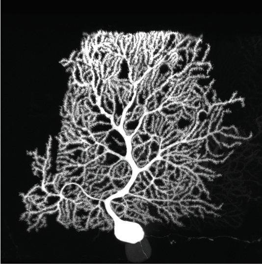
Purkinje cells are large neurons that form in the cerebellum, the small compact portion of the brain behind the cerebrum. Intricate, dendritic trees extend from each cell, forming a convoluted branching pattern that resembles a sea fan. Dysfunction of Purkinje cells has been linked to neurological symptoms, such as tremors, irregular muscle movement and hyperreactivity.
Purkinje cells’ elaborate dendrites play an integral role in neuronal communication, by receiving and integrating synaptic impulses that are coordinated by proteins scattered across the surface of the cell. Despite this integral role in neural computation, an understanding of the development of these elaborate dendrites remains elusive. A study published online in the October issue of Neuron applies a new technology to profile the proteins present on the surface of Purkinje cells to gain a better understanding of their role in dendrite development and neuronal communication.
Cell-surface proteins stick out of the cell membrane, like antennae on a building. By protruding into the surrounding environment, the proteins play a critical role in intercellular communication, like the highly orchestrated interactions between the different cell types in the nervous systems of mammals. Most techniques for studying these proteins are unable to quantify and profile the proteins in the diverse cell-type mosaic of the mammalian brain.
“Cell surface proteins are like little machines on the outside surface of cells that mediate communication between different types of cells,” said study lead author Andrew Shuster, a postdoc at Harvard University and former member of the Luo lab. “We were interested in developing a method to profile the entire coat of cell surface proteins in healthy tissue that has not been approachable by other methods.”
(more…)